Agenda
Conference Schedule
Day 1 full schedule
February 15, 2023 @ -
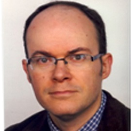
Xavier Riaud
Associate ProfessorNational Academy Of Dental Surgery
France
ABSTRACT
If we dwell upon Baron General Louis-François Lejeune’s painting entitled “The Battle of Moskova” and painted in 1822, we can notice a man standing in the corner of the canvas. The man in uniform has brown hair and is putting a bandage around an injured man’s face. If we look at it carefully, we can easily identify that this man who is practicing surgery is none other than Dominique Larrey, the chief surgeon of Napoleon’s Great Army in 1812. On September 7th, 1812, in the morning (around 10 or 11 o’clock), General Morand, who commanded the 1st infantry division of Davout’s corps, had his jaw crushed by shrapnel. Larrey treated him and the General carried on commanding his troop with gestures all through the Russian retreat.
So, what about maxillofacial surgery in the Napoleonic area?
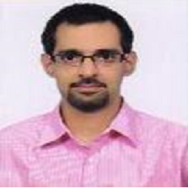
Amarjeet Gambhir
Associate ProfessorLady Hardinge Medical College & Hospital
India
ABSTRACT
Reconstruction of the maxillofacial region has always been a challenge owing to the complexity of function & aesthetics. The reconstruction of segmental mandibulectomy defects with free vascularised bone flaps is the current standard of care. A variety of donor sites, such as the iliac crest, radius, scapula & fibula have been used for this purpose with varying long-term results. Each technique has its own advantages & disadvantages. The radius & iliac crest have limited length while the scapula has limited width.
A free vascularized fibula flap was first used by Hidalgo in 1989 for the reconstruction of mandibular continuity defects and has indeed become the most popular reconstruction method nowadays. The fibula flap presents many distinct advantages, such as consistent shape, ample length & volume of bone, segmental blood supply, long vascular pedicle, distant location, low donor site morbidity, and compatibility with implant placement. The only limitation is that its height may be insufficient to restore the alveolar arch when reconstruction involves a dentate mandible, resulting in subsequent difficulty in optimal prosthetic rehabilitation with conventional dentures or osseointegrated implants. A variety of modifications have been used to overcome the height discrepancy between the native mandible and the grafted fibula. These include vertical distraction of the fibular segment as well as the use of double-barrel, one and a half-barrel, or partial double-barrel vascularized free fibular flap. All these techniques result in enhanced aesthetic & functional results. Recently, preoperative virtual surgical planning (VSP) has been used in combination with prefabricated surgical plate templates and cutting guides to make fibula flap molding & placement easier. This application of computer-assisted mandibular reconstruction (CAMR) has increased the accuracy of preoperative planning, resulting in greater surgical precision and reduction of surgical duration & postoperative complications.
To conclude, the free fibula flap because of its versatility & predictable outcome can currently be regarded as the “gold standard” for the primary or secondary reconstruction of composite oral cavity defects.
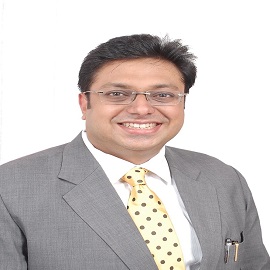
Ashish Gupta
ProfessorGupta Dental Centre - The Multispecialty Centre
India
ABSTRACT
Covid 19 is a pandemic affecting everyone across the globe. It has also changed how orthodontics is being practiced and how Aligners are helping practices across the globe. CAD CAM aligners are an acceptable method of treatment and a potent tool in the hands of a skilled Orthodontist. Although they first made their foray into Indian Markets way back in 2010 but they have really caught up the fancy of Orthodontist in India from 2015 when Invisalign made its entry into India. The presentation highlights the journey so far, the heartbreaks, the challenges, the emergence of intra oral scanners, and small little triumphs of my journey with this appliance so far in 6 years, and what the future beholds in this exciting era of Digital Orthodontics in India, especially in times of Covid 19.

Akram Belmehdi
Dental DoctorRegional center of oral care, Rabat
Morocco
ABSTRACT
The pandemic of coronavirus disease (COVID-19) is considered the biggest global health crisis for the world since the Spanish flu, also known as the 1918 flu pandemic. Driven by the SARS-CoV-2 novel coronavirus infection, the rapid spread of this disease and the related pneumonia COVID-19 are a challenge for healthcare systems in over the world, and it is a constantly evolving situation with new symptoms and prognostic factors.
SARS-CoV-2 has lately been detected in infected patient’s oral cavity; the COVID-19 outbreak is an alert that all dental and other health professionals must be vigilant in defending against the infectious disease spread.
This work summarizes an update from current medical literature about the relationship between the oral cavity and coronavirus disease by presenting some oral aspects which were detected in infected patients such as the oral lesions related to this virus and its therapeutic protocol, taste disorders, and also the diagnostic value of saliva for SARS-CoV-2.
Stereolithographic Additive Manufacturing of Ceramic Dental Crowns with Functionally Modulated Microstructures
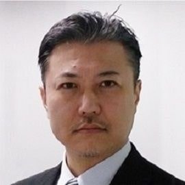
Soshu Kirihara
ProfessorOsaka University
Japan
ABSTRACT
In stereolithographic additive manufacturing (STL-AM), 2-D cross sections were created through photo polymerization by UV laser drawing on spread resin paste including nanoparticles, and 3-D models were sterically printed by layer lamination. The lithography system has been developed to obtain bulky ceramic components with functional geometries. An automatic collimeter was newly equipped with the laser scanner to adjust the beam diameter. Fine or coarse beams could realize high resolution or wide area drawings, respectively. As the row material of the 3-D printing, nanometer sized metal and ceramic particles were dispersed into acrylic liquid resins at about 60 % in volume fraction. These materials were mixed and deformed to obtain thixotropic slurry. The resin paste was spread on a glass substrate with 50 μm in layer thickness by a mechanically moved knife edge. An ultraviolet laser beam of 355 nm in wavelength was adjusted to 50 μm in variable diameter and scanned on the spread resin surface. Irradiation power was automatically changed for an adequate solidification depth for layer bonding. The composite precursors including nanoparticles were dewaxed and sintered in the air atmosphere. In recent investigations, ultraviolet laser lithographic additive manufacturing (UVL-AM) was newly developed as a direct forming process of fine metal or ceramic components. As an additive manufacturing technique, 2-D cross sections were created through dewaxing and sintering by UV laser drawing, and 3-D components were sterically printed by layer laminations with interlayer joining. Through computer-aided smart manufacturing, design, and evaluation (Smart MADE), practical material components were fabricated to modulate energy and material transfers in potential fields between human societies and natural environments as active contributions to Sustainable Development Goals (SDGs).
Yeganeh Arian
StudentBahonar Hospital
Iran
ABSTRACT
Aim :
The aim of this study was to examine the articles on the new use of engineered tissues in the treatment of problems joint temporomandibular jaw can be.
Materials and methods :
In order to carry out this study, a review of all articles sourcebooks Medline (PubMed) and Google scholar with a focus on the issue of the use of engineered tissues in the temporomandibular disorders in the period of time 1990 to 2018 about the search and investigate supposed to be.
Results:
Using the approach of engineered tissue for the different treatment of defects of temporomandibular joint disorders can be helpful is that to them out of there. The ability to build cartilage similar to cartilage naturally by way of a new provision has been. Also to help gene therapy, cell therapy, reconstruction defect of osteochondral, Ramus even part of the condyle of the left with the ability to comply with part of the left and doing function properly with the remaining part of the use of cells from stem mesenchymal (MSCs). There are factors of growth and cytokine are and provide a scaffold made of polymeric biocompatible and industries can be differentiated cells from stem mesenchymal to cells of chondrocytes and osteoblasts to cause it. Although they are tried on this is that more regeneration of muscle - Skeletal with the use of the technology and rehabilitation of cells to patients without using scaffolds.
Arnold Weiss
ProfessorTufts University School of Dental Medicine
USA
ABSTRACT
Dr. Weiss will introduce new concepts in restorative dentistry from minimally invasive care to indirect pulp therapy, and stainless steel crowns. To have a well rounded practice and encourage referrals, every practitioner needs to know how to care for children effectively. In this session you will learn how to include new procedures and get tips and secrets that you never were taught in school. Guaranteed to get many pearls in diagnosis and technique.
Goals:
1. Update on restorative dentistry
2. to be able to care for young and difficult patients
3. Get a view into minimally invasive care


